Shannon Beres, MD
Clinical Associate Professor
Neuro-ophthalmology
“I have a lot of energy, which I funnel into being an advocate for each family I see.”
My Approach
My mother was a special education teacher, so I grew up with a model of helping others. In high school I worked with children with disabilities and was a mentor for them, which inspired me to become a doctor.
I chose child neurology because I was interested in helping children with special needs, and was inspired by the family dynamics created when there's a child that has special needs.
Diseases that involve the brain can be very alarming to a family, but with additional teaching and describing how the brain works I help families understand their child's condition. I take a family-centered approach to care and believe in making decisions and developing a management plan together as a team.
I have a lot of energy, which I funnel into being an advocate for each family I see. I spend a lot of extra time going the extra mile, whether that's further diagnostic workup or making sure their care plan is fully executed. I am here for each family every step of the way.
Locations

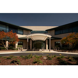
2452 Watson Court, Suite 1500
Palo Alto, CA 94303
Phone : (650) 463-9095
Fax : (650) 497-7821


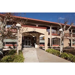
730 Welch Road, 2nd Fl
Palo Alto, CA 94304
Phone : (650) 723-0993
Fax : (650) 721-6350

730 Welch Road, 1st Fl
Palo Alto, CA 94304
Phone : (650) 800-3969
Fax : (650) 721-2884
Conditions
3rd 4th 6th Nerve Palsies
Brain Trauma
Brain Tumors
Brain and Spinal Cord Tumors
Brain, Spinal Cord and Skull Base Tumors
Cancer-Related Optic Nerve Swelling
Epilepsy Surgery
Eye Movement Abnormalities
Idiopathic Intracranial Hypertension
Myasthenia Gravis
Nystagmus
Optic Disc Atrophy
Optic Nerve Abnormalities
Optic Neuritis
Optic Neuropathy
Optic Pathway Gliomas
Papilledema
Pseudotumor Cerebri Syndrome
Septo-Optic Dysplasia
Stroke
Visual Field Defects
Work and Education
Virginia Commonwealth University School of Medicine Registrar, Richmond, VA, 6/15/2009
UCSF Dept of Child Neurology, San Francisco, CA, 8/31/2014
University of Pennsylvania Ophthalmology Fellowships, Philadelphia, PA, 7/31/2015
UCSF Pediatric Residency, San Francisco, CA, 8/28/2011
Neurology with Special Qualifications in Child Neurology, American Board of Psychiatry and Neurology, 2015
Languages
English
Publications
A zebrafish model of crim1 loss of function has small and misshapen lenses with dysregulated clic4 and fgf1b expression. Frontiers in cell and developmental biology 2025; 13: 1522094
Heterozygous deletions predicting haploinsufficiency for the Cysteine Rich Motor Neuron 1 (CRIM1) gene have been identified in two families with macrophthalmia, colobomatous, with microcornea (MACOM), an autosomal dominant trait. Crim1 encodes a type I transmembrane protein that is expressed at the cell membrane of lens epithelial and fiber cells at the stage of lens pit formation. Decreased Crim1 expression in the mouse reduced the number of lens epithelial cells and caused defective adhesion between lens epithelial cells and between the epithelial and fiber cells.We present three patients with heterozygous deletions and truncating variants predicted to result in haploinsufficiency for CRIM1 as further evidence for the role of this gene in eye defects, including retinal coloboma, optic pallor, and glaucoma. We used Clustered Regularly Interspaced Short Palindromic Repeats (CRISPR)/Cas9 to make a stable Danio rerio model of crim1 deficiency, generating zebrafish that were homozygous for a 2 basepair deletion, c.339_340delCT p.Leu112Leufs*, in crim1.Homozygous, crim1 -/- larvae demonstrated smaller eyes and small and misshapen lenses compared to controls, but we did not observe colobomas. Bulk RNA-Seq using dissected eyes from crim1 -/- larvae and controls at 72 h post fertilization showed significant downregulation of crim1 and chloride intracellular channel 4 (clic4) and upregulation of fibroblast growth factor 1b (fgf1b) and complement component 1, q subcomponent (c1q), amongst other dysregulated genes.Our work strengthens the association between haploinsufficiency for CRIM1 and eye defects and characterizes a stable model of crim1 loss of function for future research.
View details for DOI 10.3389/fcell.2025.1522094
View details for PubMedID 40114969
View details for PubMedCentralID PMC11922885
Inpatient Implementation of Portable Ocular Fundus Photography Among Neurology Residents. The Neurohospitalist 2025: 19418744251317260
Background: Nonmydriatic ocular fundus photography has been studied with demonstrated benefit in the evaluation of emergency department neurological complaints, particularly in triaging headache and focal neurological deficits. Likewise, portable fundus camera usage may be practical for inpatients with neurological complaints, although feasibility has not been studied in a neurology teaching service. Purpose: The objective of this study is to determine if a portable, nonmydriatic fundus camera could be integrated into routine clinical care by neurology inpatient housestaff at a tertiary medical center. Research Design: Housestaff were asked to obtain fundus photographs for patients with specific indications for fundoscopy. Study Sample: During a 1-month pilot period, housestaff were successfully able to upload images from 21 patients, which were reviewed by a neuro-ophthalmology attending, with input from on-call ophthalmology if desired. Results: Surveys of housestaff before (n = 13) and after (n = 12) implementation demonstrated increased confidence in camera operation and in ocular structure identification, description, and interpretation. Thematic analysis on qualitative feedback suggested benefits in clinical (improving fundus visualization, aiding in triage, sharing images with offsite staff), health systems (reducing length of stay, reducing ophthalmology consultations, reduced unnecessary testing), and educational domains (facilitating group discussions of images, sharing photographs with patients). Conclusions: Overall, inpatient portable fundus photography was shown to be feasible and effective for rapid fundus visualization for neurological inpatients, enhancing the ability to share, document, and compare examinations among neurology housestaff. Further work is needed to confirm clinical and educational benefits of portable fundus photography usage by neurology residents, as suggested by this healthcare quality improvement pilot study.
View details for DOI 10.1177/19418744251317260
View details for PubMedID 39895825
View details for PubMedCentralID PMC11780609
Author Response: Teaching Video NeuroImage: Infantile Upbeat Nystagmus as an Isolated Presentation ofCACNA1F-Related Retinal Dystrophy. Neurology 2024; 103 (12): e209667
View details for DOI 10.1212/WNL.0000000000209667
View details for PubMedID 39626124
Author Response: Teaching Video NeuroImage: Infantile Upbeat Nystagmus as an Isolated Presentation ofCACNA1F-Related Retinal Dystrophy. Neurology 2024; 103 (12): e209684
View details for DOI 10.1212/WNL.0000000000209684
View details for PubMedID 39626128
Cerebroventricular deformation and vector mapping, a topographic visualizer for surgical interventions in pediatric hydrocephalus. Journal of neurosurgery. Pediatrics 2024: 1-9
Hydrocephalus is a challenging neurosurgical condition due to nonspecific symptoms and complex brain-fluid pressure dynamics. Typically, the assessment of hydrocephalus in children requires radiographic or invasive pressure monitoring. There is usually a qualitative focus on the ventricular spaces even though stress and shear forces extend across the brain. Here, the authors present an MRI-based vector approach for voxelwise brain and ventricular deformation visualization and analysis.Twenty pediatric patients (mean age 7.7 years, range 6 months-18 years; 14 males) with acute, newly diagnosed hydrocephalus requiring surgical intervention for symptomatic relief were randomly identified after retrospective chart review. Selection criteria included acquisition of both pre- and posttherapy paired 3D T1-weighted volumetric MRI (3D T1-MRI) performed on 3T MRI systems. Both pre- and posttherapy 3D T1-MRI pairs were aligned using image registration, and subsequently, voxelwise nonlinear transformations were performed to derive two exemplary visualizations of compliance: 1) a whole-brain vector map projecting the resulting deformation field on baseline axial imaging; and 2) a 3D heat map projecting the volumetric changes along ventricular boundaries and the brain periphery.The patients underwent the following interventions for treatment of hydrocephalus: endoscopic third ventriculostomy (n = 6); external ventricular drain placement and/or tumor resection (n = 10); or ventriculoperitoneal shunt placement (n = 4). The mean time between pre- and postoperative imaging was 36.5 days. Following intervention, the ventricular volumes decreased significantly (mean pre- and posttherapy volumes of 151.9 cm3 and 82.0 cm3, respectively; p < 0.001, paired t-test). The largest degree of deformation vector changes occurred along the lateral ventricular spaces, relative to the genu and splenium. There was a significant correlation between change in deformation vector magnitudes within the cortical layer and age (p = 0.011, Pearson), as well as between the ventricle size and age (p = 0.014, Pearson), suggesting higher compliance among infants and younger children.This study highlights an approach for deformation analysis and vector mapping that may serve as a topographic visualizer for therapeutic interventions in patients with hydrocephalus. A future study that correlates the degree of cerebroventricular deformation or compliance with intracranial pressures could clarify the potential role of this technique in noninvasive pressure monitoring or in cases of noncompliant ventricles.
View details for DOI 10.3171/2024.6.PEDS24117
View details for PubMedID 39178478
Benign Ocular Flutter. The Journal of pediatrics 2024: 114229
View details for DOI 10.1016/j.jpeds.2024.114229
View details for PubMedID 39178940
DEMOGRAPHIC, CLINICAL AND IMAGING CHARACTERISTICS OF NEWLY DIAGNOSED OPTIC PATHWAY GLIOMAS ASSOCIATED WITH NF1: RESULTS FROM THE INTERNATIONAL MULTICENTER NF1-OPG NATURAL HISTORY STUDY OXFORD UNIV PRESS INC. 2024
View details for DOI 10.1093/neuonc/noae064.579
View details for Web of Science ID 001252720000545
TUMOR VOLUME IN NEWLY DIAGNOSED OPTIC PATHWAY GLIOMAS ASSOCIATED WITH NF1 (NF1-OPG): PRELIMINARY RESULTS FROM THE INTERNATIONAL MULTICENTER NF1-OPG NATURAL HISTORY STUDY OXFORD UNIV PRESS INC. 2024
View details for DOI 10.1093/neuonc/noae064.582
View details for Web of Science ID 001252720000219
Blood-born biomarkers for optic disc drusen ASSOC RESEARCH VISION OPHTHALMOLOGY INC. 2024
View details for Web of Science ID 001312227700177
Trial Design for Identifying Biomarkers of Visual Function in Optic Pathway Gliomas Secondary to Neurofibromatosis Type 1 ASSOC RESEARCH VISION OPHTHALMOLOGY INC. 2024
View details for Web of Science ID 001312227704024

Connect with us:
Download our App: