
James Gamble, MD
Professor
Pediatric Orthopedic Surgery
“It is very rewarding to help children heal and get back to doing the things they love.”
My Approach
As a pediatric orthopedic surgeon with a special interest in sports medicine, I have enjoyed fixing kids for the last 35 years. I treat orthopedic problems such as congenital anomalies, foot deformities, knee and overuse injuries, hip dislocations, and gait problems. One of my recent sources of professional pride was when a parent, who I treated as a child, brought her son in to see me because of a sports injury.
My goal is to provide personalized care that is tailored to meet the unique needs of children with orthopedic problems. I've always enjoyed working with children they have an incredible honesty and they want to get better. It's gratifying help them get back to school, back to sports, and back to a normal life.
One of the biggest changes I have enjoyed in the field has been the development of minimally invasive surgery to fix athletic injuries and fractures. Its a huge change that has resulted in safer surgery, less discomfort, and better outcomes. It is very rewarding to help children heal and get back to doing the things they love.
Locations
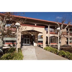
730 Welch Road, 1st Floor
Palo Alto, CA 94304
Phone : (844) 416-7846
Fax : (650) 497-8891
1195 West Fremont Avenue
Sunnyvale, CA 94087
Phone : (844) 416-7846
Conditions
ACL Tears
Adolescent Knee Pain
Clubfoot
Foot and Ankle
Gait Abnormalities
Growth Related Problems
Hip Dislocation
Knee Injuries
Low Bone Density
Orthopedics
Pediatric and Adolescent Sports Injuries
Shoulder Instability
Sports Ankle Injury
Sports Injuries by the Hip
Sports injuries
Work and Education
University of Maryland School of Medicine, Baltimore, MD, 06/30/1974
University of Maryland Hospital for Children, Baltimore, MD, 06/30/1980
University of Maryland Hospital for Children, Baltimore, MD, 06/30/1976
Johns Hopkins University School of Medicine, Baltimore, MD, 06/30/1971
University of Maryland Hospital for Children, Baltimore, MD, 06/30/1975
Orthopaedic Surgery, American Board of Orthopaedic Surgery, 1981
Languages
English
Spanish
Publications
Knee valgus and patellofemoral instability after pediatric anterior cruciate ligament reconstruction: acase report and review of the literature. Journal of medical case reports 2023; 17 (1): 212
BACKGROUND: Pediatric athletes who undergo anterior cruciate ligament reconstruction are at risk for a growth deformity if the surgery violates the physes.CASE: A 12-year-old African American boy underwent anterior cruciate ligament reconstruction using a hamstring autograft. The procedure violated the distal femoral growth plate and the perichondrial ring of LaCroix, resulting in a distal femoral lateral physeal growth arrest. Three years later, he had developed a 15° valgus deformity, an increased quadriceps angle and patellofemoral instability. He was able to return to sports after undergoing a distal femoral osteotomy to correct the valgus and medial patellofemoral ligament reconstruction to stabilize the patella.CONCLUSION: Anterior cruciate ligament reconstruction in athletes with open physes has the potential to cause distal femoral valgus deformity, an increased quadriceps angle, and subsequent patellofemoral instability.
View details for DOI 10.1186/s13256-023-03920-2
View details for PubMedID 37211594
Dostoevsky, The Brothers Karamazov, and Epilepsy. Cureus 2023; 15 (5): e38602
Fyodor Mikhailovich Dostoevsky was a brilliant nineteenth-century Russian novelist who had a seizure disorder that influenced his life and his creativity. His novels explore issues of love, faith, doubt, morality and reflect his personal experience with epilepsy. He was a keen observer of familial psychodynamics. The Brothers Karamazov (1880)was Dostoyevsky's longest and last novel, completed just a few months before his death from a pulmonary hemorrhage, most likely related to his life-long habit of cigarette smoking. In this novel, he explores the subtility of interpersonal relationships and the psychopathology within the Karamazov family and how one of the three brothers, Smerdyakov, uses psychogenic non-epileptic seizures as an alibi to get away with the perfect crime of patricide.
View details for DOI 10.7759/cureus.38602
View details for PubMedID 37168406
View details for PubMedCentralID PMC10166408
Late Presentation of a Retained Wood Foreign Body as an Expanding Soft-Tissue Mass in an Adolescent's Foot: A Case Report. JBJS case connector 2023; 13 (2)
CASE: A 12-year-5-month-old boy presented with a 3-month history of a 2 * 3-cm enlarging painful mass on the medial plantar aspect of his left foot. The radiograph was normal, but the magnetic resonance (MR) images clearly disclosed a foreign body in the shape of a toothpick that had been quiescent for 31 months. Thirty-three months after surgical removal, the patient was asymptomatic and had returned to full activity.CONCLUSION: A retained wood foreign body can present as an expanding mass, and MR is the modality of choice to image wood foreign bodies.
View details for DOI e22.00772
View details for PubMedID 37279299
Indications for and Risks Associated With Implant Removal After Pediatric Trauma. Journal of the American Academy of Orthopaedic Surgeons. Global research & reviews 2022; 6 (4)
A wide range of implants are used in the treatment of pediatric fractures, including wires, plates, screws, flexible rods, rigid rods, and external fixation devices. Pediatric bones differ from adult bones both mechanically and biologically, including the potential for remodeling. Implants used in pediatric trauma patients present a unique set of circumstances regarding indications, risks, timing of implant removal, weight-bearing restrictions, and long-term sequelae. Indications for implant removal include wire/pin fixation, when substantial growth remains, and infection. When considering implant removal, the risks and benefits must be assessed. The primary risk of implant removal is refracture. The timing of implant removal varies widely from several weeks to a year or more with the option of retention depending on the fracture, type of implant, and skeletal maturity of the patient.
View details for DOI e22.00050
View details for PubMedID 35427259
Indications for and Risks Associated With Implant Removal After Pediatric Trauma JOURNAL OF THE AMERICAN ACADEMY OF ORTHOPAEDIC SURGEONS GLOBAL RESEARCH AND REVIEWS 2022; 6 (4)
View details for DOI 10.5435/JAAOSGlobal-D-22-00050
View details for Web of Science ID 000783752200002
Remodeling of Sagittal Plane Malunion After Pediatric Supracondylar Humerus Fractures. Journal of pediatric orthopedics 2021; 41 (8): e700-e701
View details for DOI 10.1097/BPO.0000000000001912
View details for PubMedID 34397787
Radial Width of the Lateral Meniscus at the Popliteal Hiatus: Relevance to Saucerization of Discoid Lateral Menisci. The American journal of sports medicine 2021: 3635465211056661
A discoid lateral meniscus (DLM) is a congenital anomaly of the knee in which the lateral meniscus has an "O" shape and contains irregular, abnormal collagenous tissue. A DLM can cause mechanical symptoms and pain. Treatment of a symptomatic DLM is arthroscopic saucerization to reshape the meniscus to a more normal contour. Enough tissue must be removed to eliminate mechanical symptoms but not too much to create instability. The residual width of the meniscus is crucial at the popliteus hiatus because here the peripheral rim is unattached at the capsule. Reports in the literature recommend a residual width of 6 to 8 mm.The purpose of this research was to determine the width of the lateral meniscus at the popliteal hiatus in normal specimens. Our null hypothesis was that a residual width of 6 to 8 mm will be sufficient to approximate normal anatomy.Cross-sectional study; Level of evidence, 3.We made direct measurements of the radial width of the lateral meniscus from the outer rim at the popliteal hiatus to the inner edge in 19 specimens (age, 2-120 months.) We measured one 4-year-old specimen with a bilateral complete DLM. We also measured 39 digital images of specimens (age, 1-132 months) using ImageJ. Finally, we made direct arthroscopic measurements of 8 skeletally mature specimens.The average width of specimens <3 years old was 5.5 mm. The average width of the 10-year-old specimens was 12 mm. The average width of the skeletally mature specimens was 16 mm. A 4-year-old DLM specimen measured 19 mm.We rejected our null hypothesis. Direct measurements suggest that a residual width of 6 to 8 mm is insufficient for children ≥8 years old. A width of at least a full centimeter approximates the normal for 8-year-olds and at least 15 mm for adolescents.
View details for DOI 10.1177/03635465211056661
View details for PubMedID 34780308
Late Stabilization of Developmental Dysplasia of the Hip Without Treatment: A Case Report. JBJS case connector 2020; 10 (4)
CASE: Approximately three-quarters of neonates with unstable hips will spontaneously stabilize without treatment in the first few weeks of life. This report presents the long-term follow-up of an infant with developmental dysplasia of the hips that stabilized at an older age and without any orthopaedic treatment.CONCLUSIONS: Factors contributing to the spontaneous stabilization in this case included the patient's self-selected lower extremity position of comfort with hips flexed, abducted, and externally rotated; her delayed walking; and her light body weight.
View details for DOI 10.2106/JBJS.CC.20.00294
View details for PubMedID 33512920
Bilateral Medial Patella Sleeve Fractures in a Child: A Case Report. JBJS case connector 2020; 10 (2): e1900533
CASE: An 8-year-old girl presented with a displaced right medial patella sleeve fracture. She underwent open reduction and suture fixation. Three years later, she presented with a left medial patella sleeve fracture that was less displaced than on the right. This was treated with immobilization and structured rehabilitation. She was able to return to full activity with normal radiographs at the final follow-up.CONCLUSION: Patella sleeve fractures are rare. We report a unique case of bilateral medial patella sleeve fractures in an otherwise healthy child in which one side was treated operatively and the other was treated nonoperatively.
View details for DOI 10.2106/JBJS.CC.19.00533
View details for PubMedID 32649152
Type VII All-Epiphyseal Fractures of the Lateral Malleolus and the Origin of Subfibular Ossicles. Journal of pediatric orthopedics 2020
A subfibular ossicle (SO), also known as an os subfibulare, is present in ∼1% of the general population. Two theories have been proposed to explain the origin of SOs: (1) as a failure of fusion of a secondary center of ossification; (2) as a posttraumatic sequela. This report offers prospective, longitudinal radiographic evidence for the formation of SOs as a posttraumatic sequela of type VII transepiphyseal fractures of the lateral malleolus in children.This Institutional Review Board-approved study was performed at a tertiary care pediatric hospital from March 2012 to April 2019. The study group included 37 children with a type VII fracture of the lateral malleolus and a minimum follow-up of 6 months.Twenty-one children (57%) healed their fracture. Sixteen children (43%) went on to form SOs. The most common location for the fractures was the distal third of the epiphysis, and the most common fracture type forming SOs was a sleeve avulsion fracture. Four of the children forming SOs have had surgery to address pain and recurrent sprains.Overall, 43% of children who sustained a type VII fracture of the lateral malleolus went on to form SOs, giving support to the posttraumatic theory of origin. Sleeve avulsion fractures have the greatest chance of forming SOs. So far, 4 of the 16 children forming SOs have undergone surgery for ankle pain and recurrent sprains.Level II.
View details for DOI 10.1097/BPO.0000000000001638
View details for PubMedID 32675577

Connect with us:
Download our App: